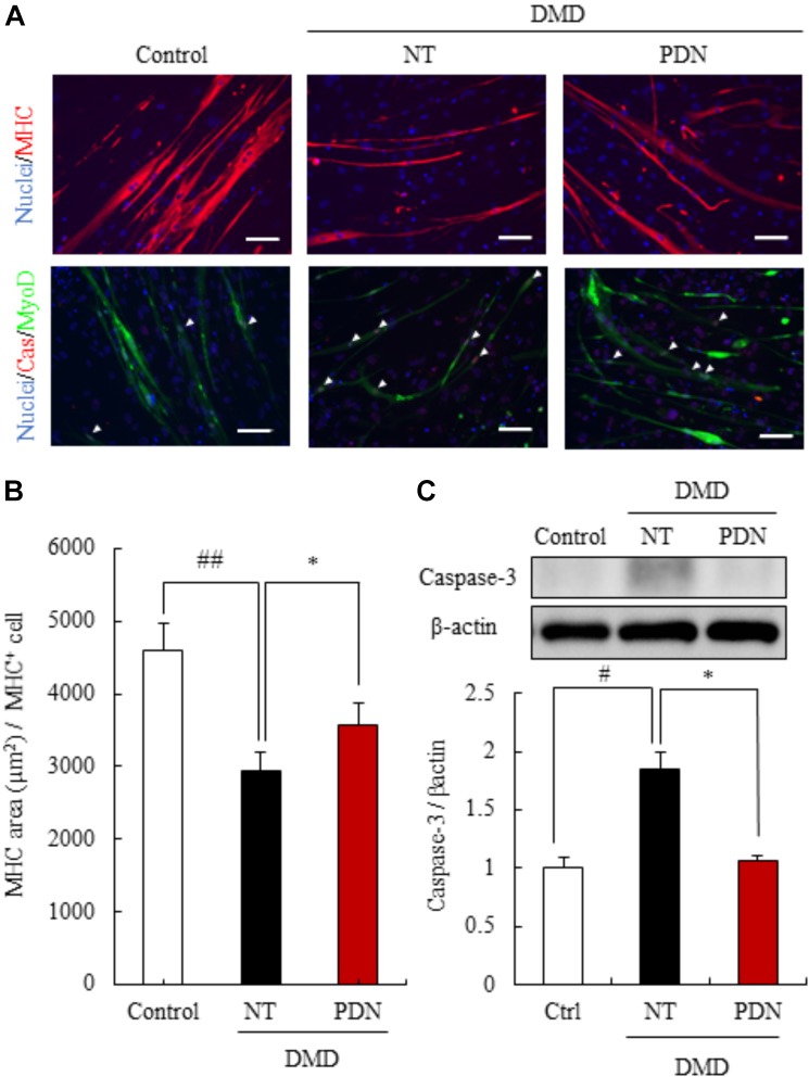
FIGURE 4.
Efficacy of prednisolone in reversing DMD pathology. (A) Immunostaining using skeletal muscle marker (myosin heavy chain, MHC), and apoptosis marker (cleaved caspase-3, Cas). Prednisolone (PDN) restored decreased MHC area and increased apoptosis cells. Scale bars = 100 μm. (B) Quantification of the MHC area of myotubes from control individual, non-treated myotubes from DMD (NT), and prednisolone-treated myotubes from DMD (PDN). Data are shown as means ± SEM (n = 4). (C) Western blot analysis of cleaved caspase-3 in control individual (Ctrl), NT, and PDN. Data are shown as means ± SEM (n = 5). #p < 0.05 vs. Ctrl and ∗p < 0.05 vs. NT (Student’s t-test), ∗p < 0.05 vs. NT (Student’s t-test).

