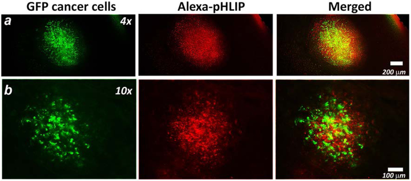
Figure 6. Targeting of submillimeter metastatic lesions in lungs.
4T1-GFP cells were injected subcutaneously in the mammary pad of the mouse. After 3 weeks, the primary tumor metastasized in the lungs. The AF546-Var3 was given as a single i.v. tail vein injection. At 4 hrs p.i. animals were euthanized, the lungs were excised and imaged immediately under fluorescent microscope. The representative fluorescent images obtained at different magnifications are presented. The GFP (green) and AF546-Var3 (red) fluorescence and their overlay are shown. The experiment was repeated on six animals and total 16 metastatic lesions were detected.

