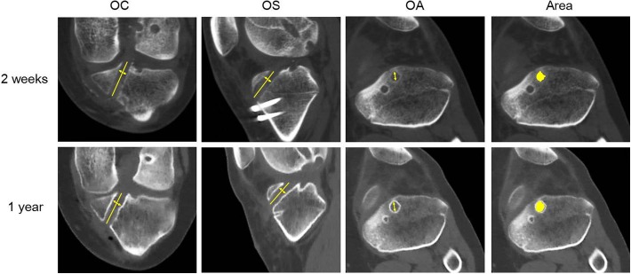
Figure 5.
Multiplanar reconstruction images from computed tomography scans of the tibia at 2-week (top row) and 1-year (bottom row) follow-up. The femoral anteromedial tunnel showed a diameter at 10 mm from the intra-articular outlet of the femoral tunnels (line with arrows). OA, oblique axial; OC, oblique coronal; OS, oblique sagittal. Cross-sectional area represented by yellow circle.

