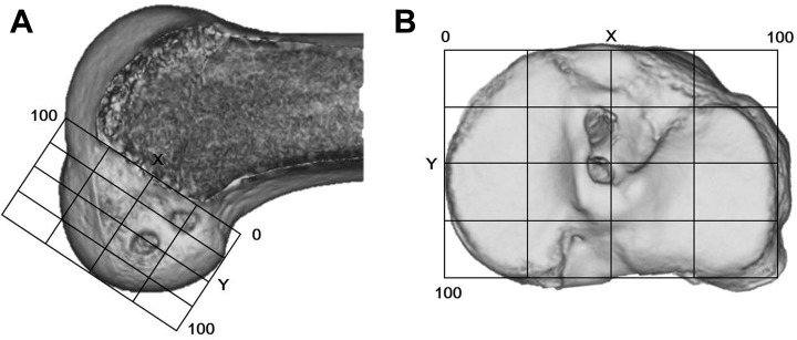
Figure 6.
The quadrant method to evaluate the position of the femoral and tibial tunnels. Evaluation of each tunnel outlet with standard and 3-dimensional computed tomography (3D CT): (A) To evaluate the center location of each femoral tunnel outlet, we drew the X and Y coordinate system on the 3D CT scan so that we could obtain a correct lateral view of the femoral condyle and determine the Blumensaat line. (B) To evaluate the center location of each tibial tunnel outlet, we drew the X and Y coordinate system on a standard CT scan.

