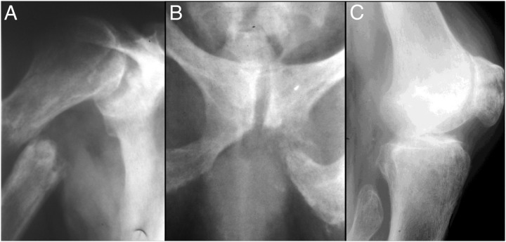
Figure 3.
Radiographs after falls and fractures. At age 55 years, radiographs showed increased osteopenia and more prominent trabecular coarsening in association with: medially displaced right proximal humeral diaphyseal fracture inferior to the surgical neck and through an area of marked focal “osteolysis” (A); fracture of the inferior left ischiopubic ramus, again through an area of focal bone lysis (B); and transverse right patellar fracture with widened irregular margins, again consistent with preexistent focal osteolysis at the location of the fracture (C). All three fractures above have irregular margins with areas of focal osteolysis. Their appearance suggests, therefore, that they are pathological fractures that occurred through preexisting lesions (likely pseudofractures). Not illustrated were an oblique fracture through the right midulnar diaphysis, oblique fracture through the right distal femoral diaphysis around his long-stem total hip prosthesis, minimally displaced transverse fractures in the right distal femoral metaphysis, and a fracture in the proximal tibial metaphysis.

