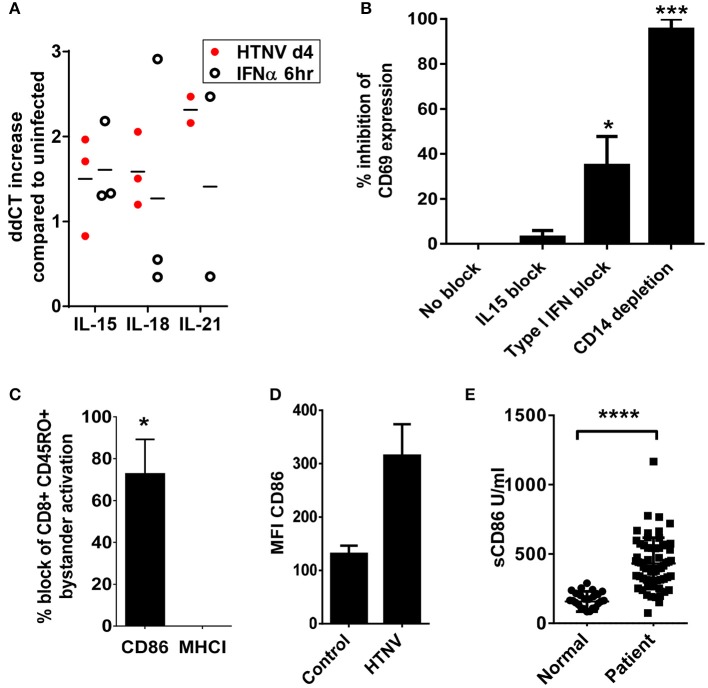
Figure 8.
Dependency of hantavirus-induced bystander activation on costimulatory CD86 molecules. (A) Immature DCs were infected with HTNV at a MOI of 1.5 and incubated for 4 days or exposed to IFN-α for 6 h at 2,000 U/ml before being harvested. Subsequently, cellular RNA was isolated and the number of indicated cytokine-encoding transcripts quantified by qPCR according to the delta-delta-Ct (ddCt) method. (B) PBMCs treated with anti-IL15 (20 μg/ml) or anti-IFN-α (20 μg/ml) and PBMCs depleted of CD14+ cells were exposed to HTNV at a MOI of 1.5 for 4 days before CD69 expression on CD8+ cells was measured by cytofluorimetric analysis. Results are derived from three independent experiments, error bars represent the mean ± SEM (*p < 0.05, ***p < 0.001, 1 way ANOVA test with Bonferroni correction). (C) PBMCs treated with anti-CD86 or anti-MHC (both 10 μg/ml) were exposed to HTNV at a MOI of 1.5 for 4 days before CD69 expression on CD8+ CD45RO+ cells was determined by cytofluorimetric analysis. Error bars represent the mean ± SEM (*P < 0.05, Student's t-test). (D) PBMC infected with MOI 1.5 of TULV or HTNV were analyzed 3 days post infection for the expression of CD86 on the surface of CD14+ cells. (E) Sera from normal healthy individuals or convalescent hantavirus-infected patients were tested by ELISA for levels of sCD86. Error bars represent the mean ± SD (****p < 0.0001, paired Student's t-test).

