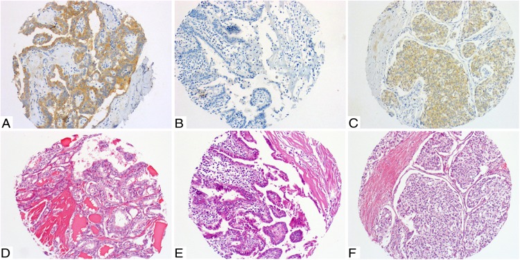
Figure 1.
A and D, Classic PTC, mutated for BRAF V600E. A, 3+ diffuse homogeneous immunostaining with VE1. D, Corresponding H&E staining. B and E, Classic PTC, wild type for BRAF V600E. B, 0 to 1+ immunostaining with VE1. E, Corresponding H&E staining. C and F, Poorly differentiated carcinoma mutated for BRAF V600E. C, 2+ diffuse homogeneous immunostaining with VE1. F, Corresponding H&E staining.

