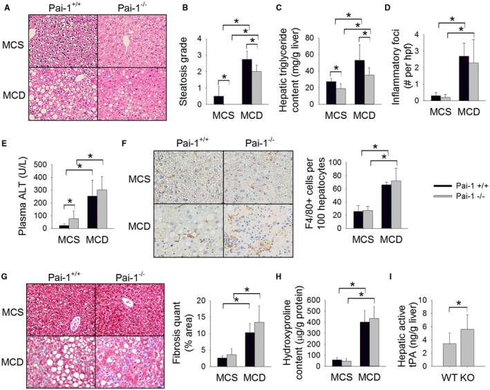
Figure 2.

Pai‐1 deletion attenuates MCD diet‐induced hepatic steatosis but does not prevent hepatic inflammation or fibrosis. (A) H&E‐stained liver sections, (B) steatosis grade, (C) hepatic triglyceride content (mg/g liver), (D) number of inflammatory foci on H&E sections per hpf, (E) plasma ALT (U/L), (F) F4/80‐stained liver sections with quantification (# cells/100 hepatocytes), (G) Masson’s trichrome‐stained liver sections with quantification of trichrome staining, (H) hepatic hydroxyproline content (µg/g protein), and (I) hepatic level of active tPA (ng/g liver) in Pai‐1 –/– and Pai‐1+/+ mice fed an MCD or MCS (control) diet for 8 weeks. Values are expressed as mean ± SD; n = 7‐8; *P < 0.05. Abbreviations: KO, knockout; WT, wild type.
