Figure 1.

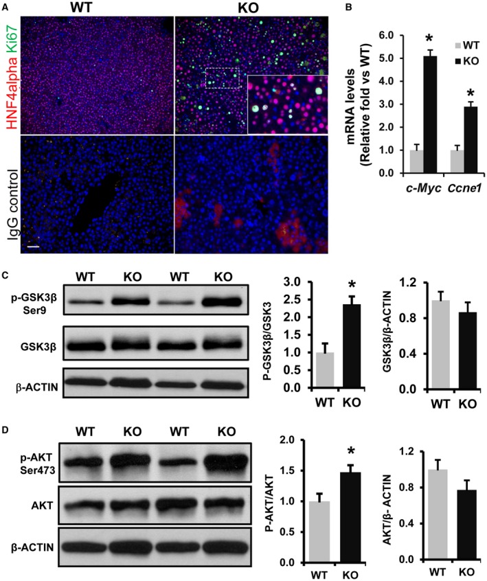
Increased cell proliferation and activation of WNT signaling in 3‐week‐old Phb1 KO livers. (A) Paraffin‐embedded liver sections from 3‐week‐old Phb1 KO mice and WT controls were subjected to co‐immunofluorescence staining for proliferation marker Ki67 (green) with hepatocyte marker HNF4alpha (red) and control IgG (staining control). Nuclei are stained with DAPI (blue). Images represent three independent staining on four different KO and WT livers. Scale bar, 25 μm. (B) Increased expression of WNT target genes in Phb1 KO liver. Total RNA was prepared from 3‐week‐old WT and Phb1KO livers and subjected to qPCR as described in Materials and Methods. Relative expressions of c‐Myc and Ccne1 were compared with WT controls. Results represent mean ± SEM from n = 7 mice in each group; *P < 0.01 versus WT. Total protein was extracted from 3‐week‐old Phb1 KO and WT livers and immunoblotted for (C) p‐GSK3betaSer9, total GSK3beta, and beta‐ACTIN and (D) p‐AKTSer473, total AKT, and beta‐ACTIN. Representative blots are shown. Protein band intensity was quantified by densitometry analysis by ImageJ software (NIH). The ratio between normalized phospho/total protein band intensities was calculated, then mean activation fold was represented over WT. Results represent mean ± SEM from n = 7 mice in each group; * P < 0.05 versus WT. Abbreviations: c‐Myc, Myc Proto‐oncogene; DAPI, 4´,6‐diamidino‐2‐phenylindole.
