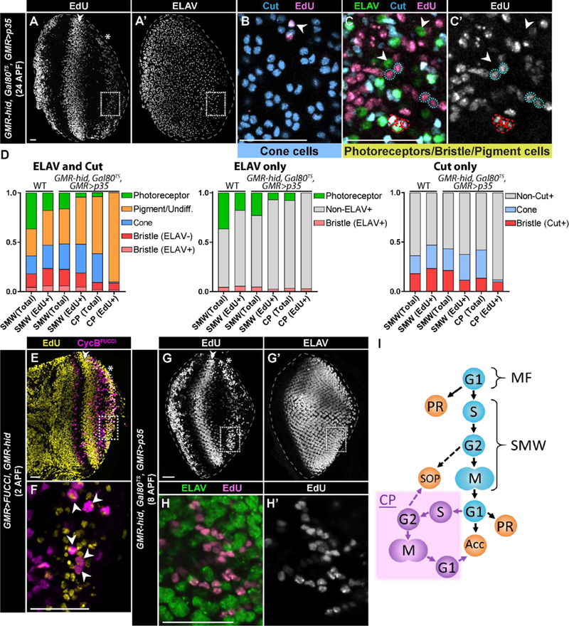
Fig. 4: Compensatory proliferating cells contribute to non-photoreceptor, accessory cells types in the retina.

(A-C’) GMR-hid, Gal80TS, GMR>p35 retina at 24hrs APF stained for EdU incorporation (A), ELAV (A’,C), and Cut (B,C) (A, B, and C from different retinas). Retinas were derived from 3rd instar larvae fed with EdU for 10 hours. The SMW (double arrowhead) and CP (asterisk) are visible in A. (B) Magnification (dotted box in A, A’) of an apical section of the CP wave displays Cut expressing cone cells (blue) that are EdU+. (C,C’) Magnification of a basal section of the CP wave displays EdU+ (pink), Cut-expressing bristle groups (red dashed circles; Cut, blue; neurons express ELAV (green)) and pigment cells (blue dotted circles; Cut/ELAV-). ELAV expressing photoreceptors (Cut-) are EdU-(arrowheads). Note that the number of photoreceptors in these retinas is reduced relative to wild type due to the continual expression of hid. (D) Quantification of the types of EdU+ pupal retinal cells after feeding EdU to 3rd instar larvae for 10 hours. Retinas were stained for ELAV and Cut, only ELAV, or only Cut. Data are presented as a percentage of the total (i.e. 1.0). SMW (Total) and SMW (EdU+) for WT were calculated based on the numbers of cells in an ommatidium (see Methods). One retina was counted per graph for SMW in GMR-hid, Gal80TS, GMR>p35; CP analysis was performed on n=5 retinas (ELAV and Cut), n=2 retinas (ELAV only), and n=2 retinas (Cut only). At least 50 EdU+ cells and 250 total cells were counted per retina. Pigment cells were identified by lack of expression of ELAV or Cut; this population may include undifferentiated (Undiff.) cells as well. (E-F) GMR>FUCCI, GMR-hid retina at ~2hrs APF derived from 3rd instar larvae fed with EdU for 10 hours and stained for EdU incorporation. Box in E,E’ indicates area of magnification in F, where a subset of cells that were EdU-labeled in the CP wave express CycBFUCCI (arrowheads). (G,G’) GMR-hid, Gal80TS, GMR>p35 retina at ~8hrs APF derived from 3rd instar larvae fed with EdU for 10 hours and stained for EdU incorporation (G) and ELAV (G’). (H,H’) Magnification (dotted box in G) demonstrates that ELAV expressing photoreceptors (green) are not labeled with EdU (pink in H, gray in H’). Scale bars=20 µM. (I) Schematic of the relationship between cell cycle phase (G1,S,G2,M) and precursor cell fate in damaged GMR-hid larval retinas. SOP denotes sensory organ precursor cells that give rise to the interommatidial bristle group of cells; PR denotes photoreceptor; Acc denotes accessory cell. Purple denotes compensatory proliferation (CP). MF, morphogenetic furrow. SMW, second mitotic wave.
