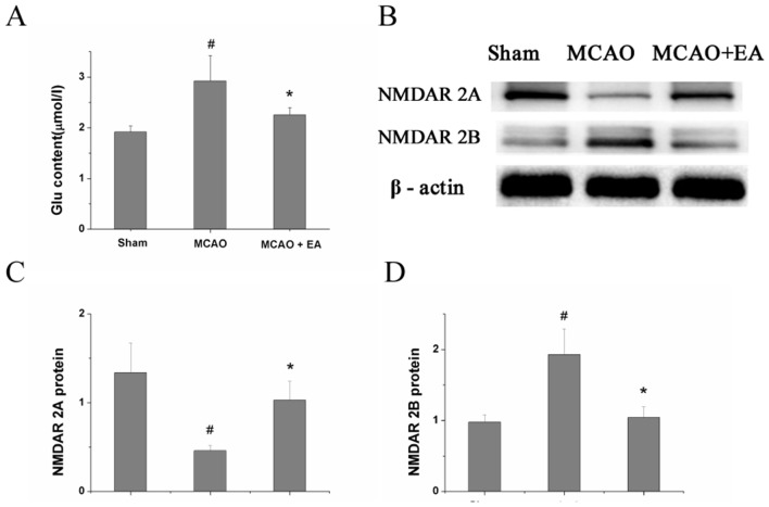
Figure 3.
Effect of EA on Glu content and expression of NMDAR2A and NMDAR2B protein. (A) Glu content in the CA1 area of the hippocampus in each group, measured by ELISA. (B) Protein expression of NMDAR2A and NMDAR2B in the CA1 area of the hippocampus in each group, examined by Western blotting. (C,D) Quantitative indices of protein expression of NR2A and NR2B, derived from figure 2B. #P<0.05 versus control group; *P<0.05 versus MCAO group. EA, electroacupuncture; Glu, glutamate; MCAO, middle cerebral artery occlusion; NMDAR2A, N-methyl-D-asparticacid receptors 2A; NMDAR2B, N-methyl-D-asparticacid receptors 2B.

