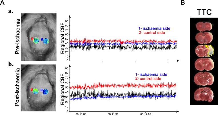
Figure 1.
Establishment of the middle cerebral artery occlusion model. Brain imaging was captured using a dynamic laser speckle technique to monitor the regional cerebral blood flow (CBF) in the skull cranial window pre-ischaemia (Aa) and post-ischaemia (Ab). TTC staining shows development of an extensive lesion in the lateral cortex (indicated by yellow arrow in (B)). TTC, 2,3,5-triphenyltetrazolium chloride.

