
Fig. 3.
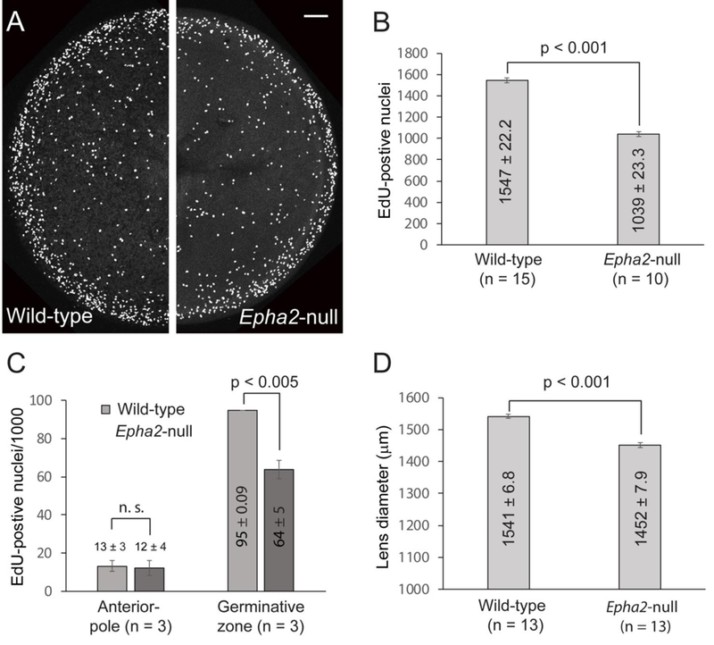
Fluorescence imaging of S-phase epithelial cell nuclei in the mouse lens (P8). (A) EdU labeling of intact wild-type (left panel) and Epha2-null (right panel) lenses showing the increased concentration of S-phase cell nuclei in the germinative zone at the lens equator (outer) compared with the anterior pole region (central). Scale bar: 100 μm. (B, C) Both the raw global count (B) and the normalized count/1000 nuclei (C) of EdU-positive nuclei in the Epha2-null lens were significantly less than wild-type. (D) The equatorial dimeter of the Epha2-null lens was also significantly less than wild-type consistent with early lens growth inhibition.