


Fig. 6.
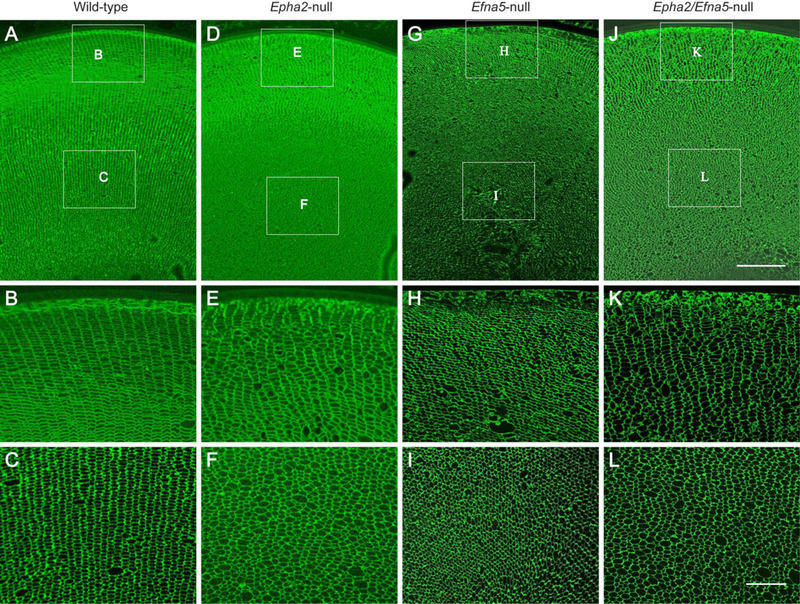
Immuno-localization of AQP0 in mouse lens sections (P30). Coronal section (crossoptical plane) view of the wild-type lens (A-C), Epha2-null lens (D-F), Efna5-null lens (G-I), and Epha2/Efna5-null lens (J-L) at P30 labeled with AQP0 antibody. Note that the radial columns of hexagonal fiber cells in the outer cortex (B) and inner cortex (C) of the wild-type lens are variably disturbed in the outer and inner cortex regions of the Epha2-null lens (E, F), Efna5-null lens (H, I), and Epha2/Efna5-null lens (K, L). Scale bar: 100 μm (A, D, G, J), 30 μm (B, C, E, F, H, I, K, L).