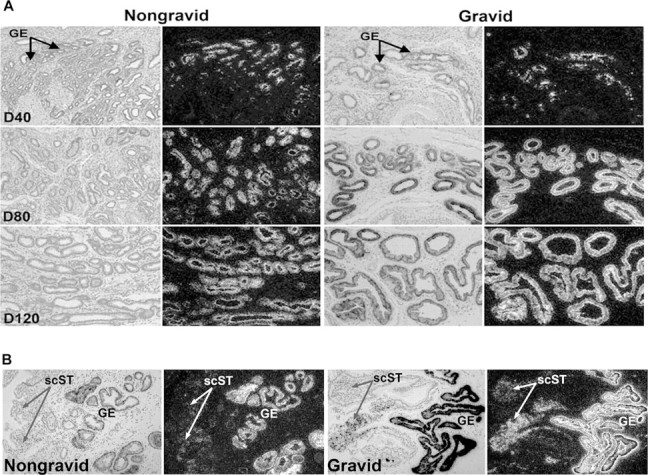
Fig. 3.

In situ hybridization analysis of SPP1 mRNA in uteri collected from unilaterally pregnant ewes. A) SPP1 mRNA in glandular epithelium (GE) on Gestational Days (D) 40, 80, and 120. B) SPP1 mRNA in stratum compactum stroma (scST) on Day 80. Corresponding brightfield and darkfield images of representative endometrial cross sections are shown. A section hybridized with radiolabeled sense cRNA probe shown in Figure 6 serves as a negative control. Width of each field is 940 μm.
