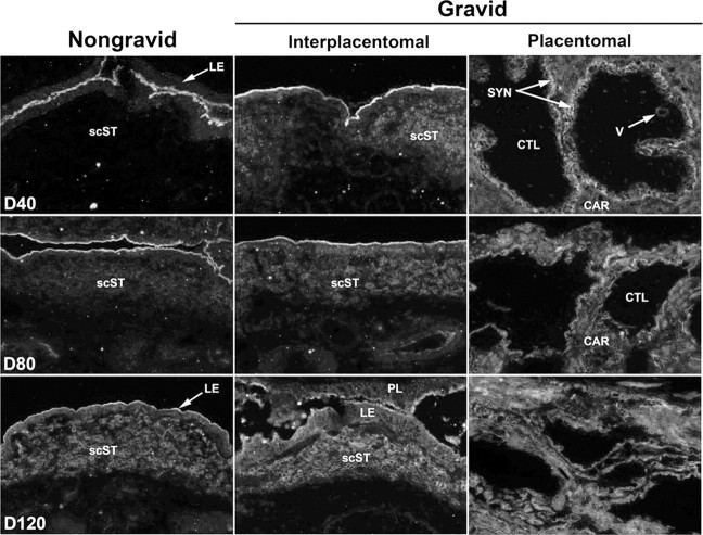
Fig. 4.
Immunofluorescence localization of SPP1 (polyclonal rabbit anti-human SPP1 IgG [LF-123 + LF-124]) in frozen sections of uteri from nongravid and gravid horns collected from unilaterally pregnant ewes on Gestational Days (D) 40, 80, and 120. Rabbit IgG (IgG) serves as a negative control and is shown in Figure 8. scST, Stratum compactum stroma; LE, luminal epithelium; PL, placental membranes; CTL, cotyledon; CAR, caruncle; SYN, syncytia; V, blood vessel. Width of each field is 940 μm.

