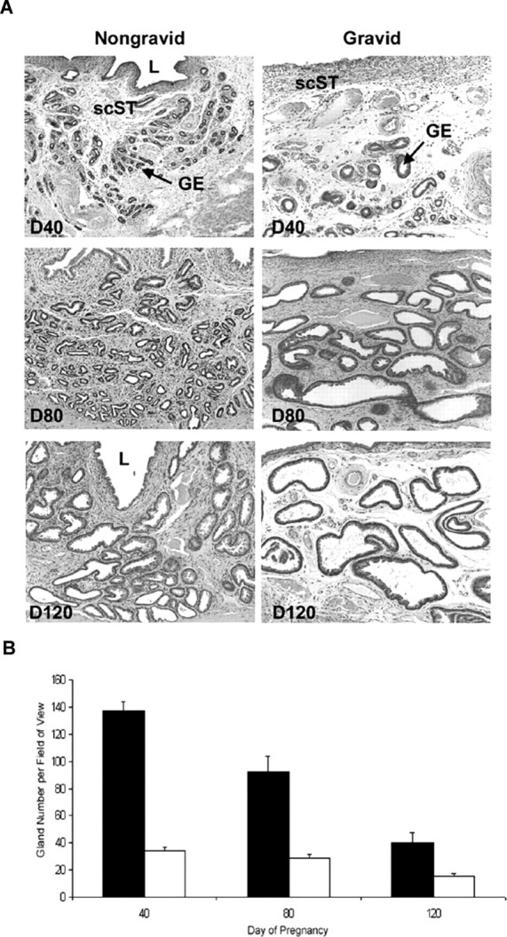
Fig. 5.

A) Representative photomicrographs of hematoxylin and eosin-stained uterine cross sections of nongravid and gravid uterine horns from unilaterally pregnant ewes collected on Gestational Days (D) 40, 80, and 120 are shown. L, Lumen; GE, glandular epithelium; scST, stratum compactum stroma. Width of each field is 940 μm. B) Quantification of uterine gland number per 940-μm field of view. Fewer glands were visible per field in gravid (white bars) than nongravid (black bars) uterine horns (P < 0.001) throughout gestation.
