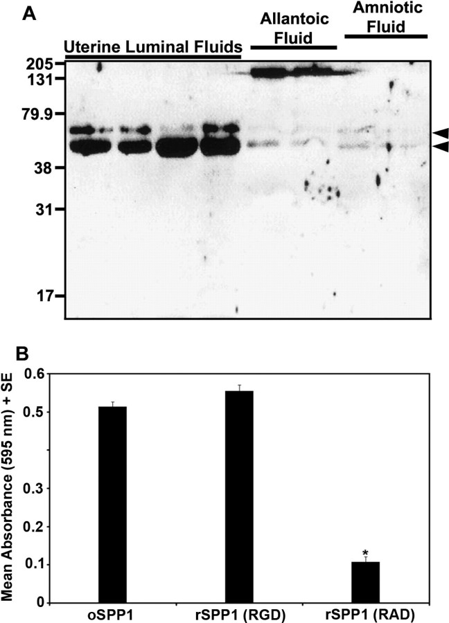
Fig. 7.
A) Western blot analysis of SPP1 in uterine luminal secretions recovered from nongravid uterine horns and from allantoic and amniotic fluids of conceptuses in the gravid uterine horn of unilaterally pregnant ewes on Gestational Day 120. Each lane represents a sample from a different ewe. Immunoreactive SPP1 was detected using a cocktail of polyclonal rabbit anti-hSPP1 IgG (LF-123 + LF-124). Positions of prestained molecular weight standards (kDa) are indicated. Arrowheads denote SPP1 fragments. B) SPP1 promotes attachment of ovine trophectoderm cells. Adhesion assays were conducted with rSPP1 (RGD), rSPP1 (RAD), or ovine uterine luminal SPP1 (oSPP1). Data represent absorbance values (595 nm) + SEM (n = 3 wells per data point). Attachment did not differ between oSPP1 and rSPP1 (RGD) (P > 0.10), but was significantly less using rSPP1 (RAD) than oSPP1 or rSPP1 (RGD) (*P < 0.01).

