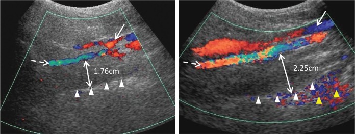
Figure 2. Sonographic measurement of lateral parapharyngeal wall thickness.
The solid line and dashed line arrows indicate the internal jugular vein and the internal carotid artery visualized by Doppler imaging, respectively. The lateral wall of pharynx is represented by the echogenic interface (white triangles). The double-headed arrow indicates the distance between the internal carotid artery and the echogenic surface of pharynx, representing the thickness of lateral parapharyngeal wall. The yellow triangles indicate the color vibration artefacts caused by the motion of the lateral wall of pharynx.

