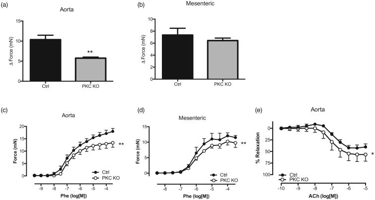
Figure 5.
Protein kinase Cα knock out mice have reduced vascular contractility. Relaxation responses to potassium chloride (120 mmol) were performed in aorta (a) and mesenteric arteries (b) from both protein kinase C knock out and control mice. Values shown are expressed as the change in force (mN). Data are represented as mean ± SEM; n = 4. **P < 0.01, Rmax values of protein kinase C knock out vs. Control. Concentration response curves were performed to phenylephrine (10 μmol/l) in aorta (c) and mesenteric arteries (d) from both protein kinase C knock out and control mice. Values shown are expressed as the change in force (mN). (e) Concentration response curves to acetylcholine were performed in phenylephrine-precontracted aorta. Relaxation responses were calculated relative to the contraction elicited by phenylephrine. Data are represented as mean ± SEM; n = 4. **P < 0.01, Rmax values of protein kinase C knock out vs. control.

