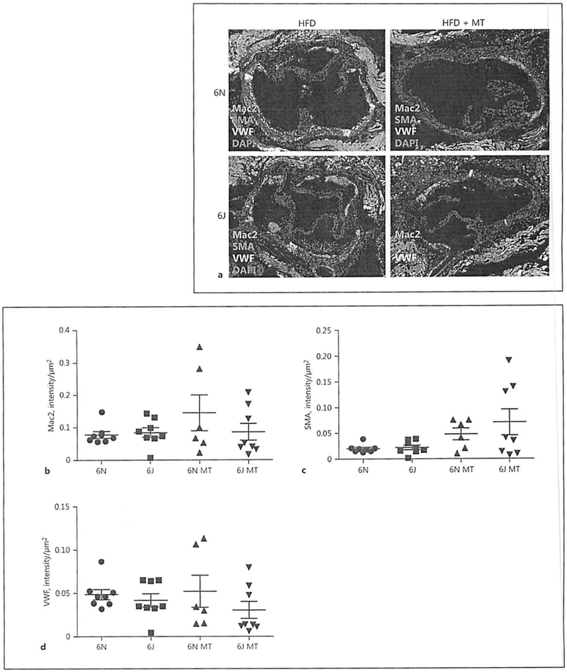
Fig. 6.

Cellular composition of aortic root plaques is similar among treatment groups. Male C57BL/6N and C57BL/6J mice were treated with AAV8-PCSK9 (3 × 10¹⁰ vector genomes) for 2 weeks and then subjected to a high-fat diet (HFD) for 8 weeks. A subset of animals was co-treated with MitoTEMPO (MT; 0.8 mg/kg/day) for the final 4 weeks of the experiment. a Representative immunofluorescence images visualizing macrophage area (Mac2 positive, green), smooth muscle (smooth-muscle actin, SMA positive, red), and endothelial (von Willebrand factor positive, VWF, white) localization within aortic root plaques. Quantification of macrophage (b), smooth muscle (c), and endothelial area (d) is provided. The scale bar is 200 μm at the ×20 magnification. Values are means ± SEM of 6–8 animals.
