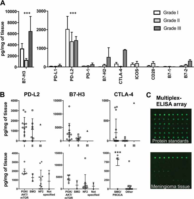
Figure 1.

Immune checkpoint protein expression in meningioma tissues according to WHO grade and genetic subtype. (A) Quantities of 10 common immune checkpoint proteins in meningioma tissues according to WHO grade (n = 22). Amounts of B7-H3 and PD-L2 proteins are significantly greater than all other proteins analyzed. (B) Expression of PD-L2, B7-H3 and CTLA-4 for each case (n = 20) is quantified according to WHO grade and gene mutation subtype. (C) Representative scans of a multiplex ELISA array for a single protein standard used in the generation of a protein quantification standard curve and also a meningioma tissue protein sample. *** P-value < 0.001. Bars represent mean protein quantity in pg/mg of meningioma tissue. Error bars represent SEM.
