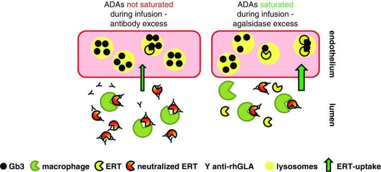
Figure 5.
Schematic model of ERT and neutralizing antibodies during infusion. If antibodies are present, they neutralize ERT activity by binding. In addition, IgG-tagged agalsidase will be internalized and digested by macrophages. If more antibodies than ERT are present (antibody excess/agalsidase deficit), this results in a decreased cellular Gb3-clearance (left). If ERT overcomes antibody titers (agalsidase excess), more ERT can enter lysosomes of target cells (right).

