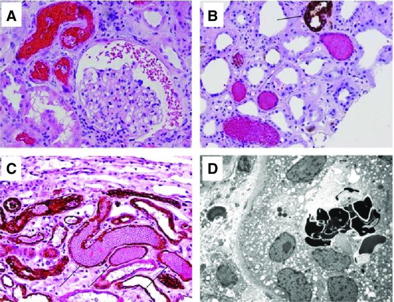
Figure 1.
Typical renal biopsy findings in warfarin-related nephropathy. Red blood cells (RBCs) in different compartments of the kidney in patients on warfarin therapy with acute kidney injury. (A) Numerous RBCs and RBC occlusive casts were noticed in tubules and Bowman space. (Hematoxylin and eosin stain; original magnification ×200). (B) Immunohistochemical stain for Tamm-Horsfall protein shows that most RBC casts do not contain Tamm-Horsfall protein. (Arrow) Positively-stained thick ascending loop of Henle. (C) Immunohistochemical stain for cytokeratin AE1/AE3 (arrows, dark brown) highlights distal tubules with occlusive RBC casts. (Counterstain with hematoxylin/eosin; original magnification ×200). (D) Dysmorphic RBCs were noticed in several tubules by means of electron microscopy. (Uranyl acetate, lead citrate stain; original magnification ×3000).

