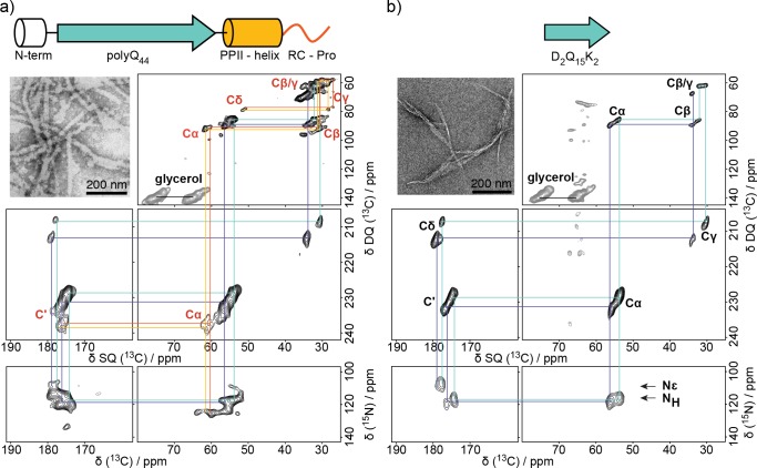
Figure 2.
13C–13C and 13C–15N correlation spectra of (a) Q44-HttEx1 and (b) D2Q15K2 fibrils. Two types of Gln that compose the polyQ core are annotated in purple and cyan, while polyproline helix and random coil prolines are annotated in red and orange, respectively. TEM micrographs of the fibril samples are shown as insets. 13C assignments for oligoproline and polyQ regions are labeled in red and black in panels a and b, respectively. Only one set of assignments is shown in panels a and b for clarity.

