
Figure 3.
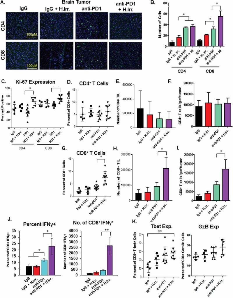
Abscopal effects induced by radiation and anti-PD1 combination therapy are associated with increased anti-tumor immunity. A. At the conclusion of the previous experiment, brain tissue was sectioned and T cell markers stained for immunofluorescence microscopy. B. Quantification of the number of cells in at least four independent fields for each treatment group. C. Flank tumor tissue was harvested and dissociated by collagenase/hyaluronidase digestion and immune cell markers were stained for readout and quantification by flow cytometry. Ki-67 expression was assessed on the indicated T cell subsets by permeabilization and staining followed by readout by flow cytometry. D-I. T cell subset percentages in non-irradiated flank tumor tissue were compared to total cell recovery to determine total number of T cells and ratio of CD8T cells to regulatory CD4T cells (Treg). CD4+/CD8+ percentages in D. and G. are of total CD45+ cells. J. CD8+ T cells from non-irradiated flank tumors were restimluated ex vivo with plate-bound anti-CD3 and anti-CD28 antibodies followed by intracellular cytokine staining for interferon-gamma (IFNγ) staining. K. Expression of the indicated markers of cytotoxic T cell function on CD8+ T cells from non-irradiated flank tumor tissue were stained following permeabilization and read-out by flow cytometry. *p ≤ 0.5 and **p ≤ 0.01 for indicated comparisons between anti-PD-1 and anti-PD-1 plus head irradiation treatment groups according to a student’s t test.