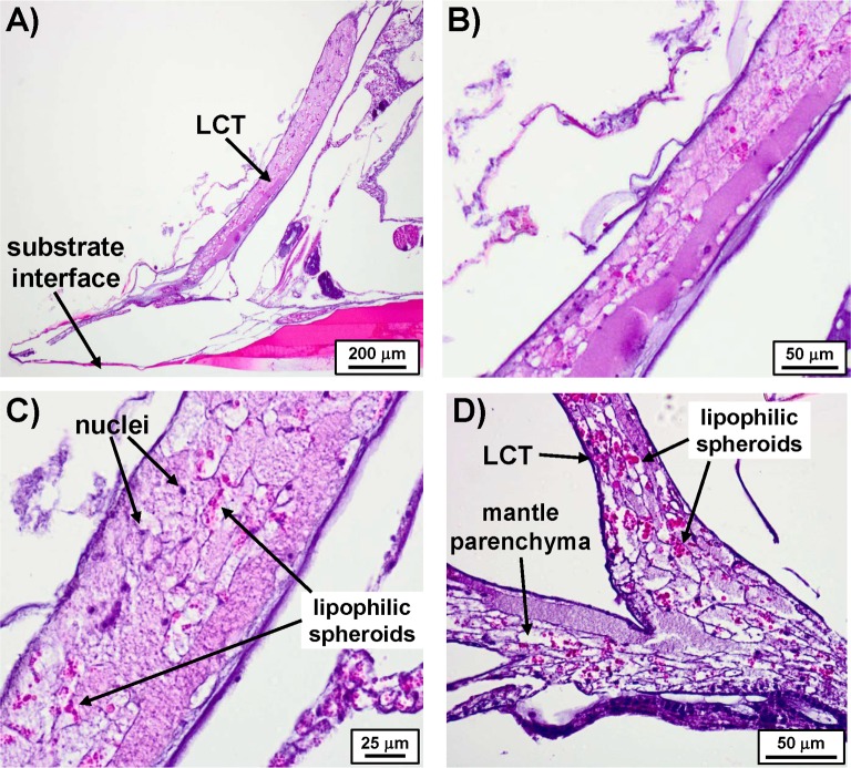
Fig 5. Sagittal sections of LCT stained with H&E.
A) Entire view of LCT in relation to the edge of A. amphitrite. B) Magnified image of A) showing the various textures and staining profile. C) Another zoomed image of A) highlighting the presence of nuclei and clusters of lipophilic spheroids. D) Sagittal section showing connection between the LCT and the mantle parenchyma.

