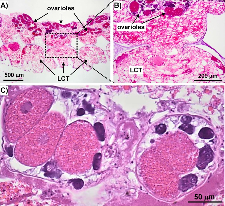
Fig 6. H&E stained transverse A. amphitrite sections.
A) Low magnification image of LCT and sub-mantle tissue containing the ovaria. B) Higher magnification image of A) highlighting continuity between LCT and sub-mantle tissue. C) Image of two ovaria showing oogenesis at different developmental stages.

