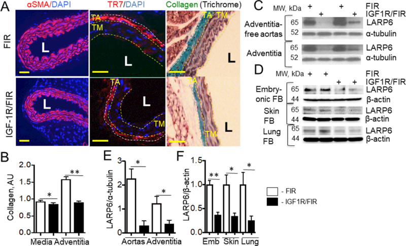
Figure 2. IGF-1R deficiency decreased vascular collagen (A, B) and downregulated LARP6 (C-F).

A, Cross-sections of descending aorta were stained for αSMA (SMC marker), TR7 (FB marker) and DAPI or stained for collagen (Trichrome) (n=6/group). LARP6 expression was quantified in adventitia-free aortas, adventitia (C, E) and in embryonic, skin and lung fibroblasts isolated from mouse tissues (D, F) (n=4/group). Yellow line, tunica media (TM), white line, tunica adventitia (TA). L – lumen, *P<0.05, **P<0.01. Scale bar, 10um.
