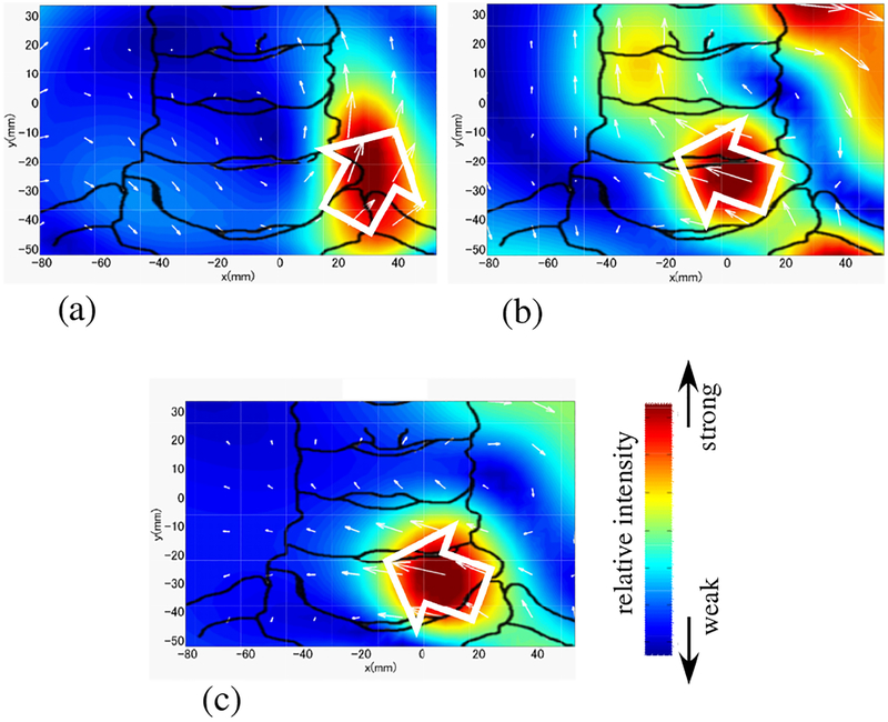
Figure 14:
Results of source reconstruction when a stimulus applied to the subject’s left median nerve. (a) Source image at the latency of 5.8 ms obtained from the original sensor data in Fig. 12(a). (b) Source image at the latency of 5.8 ms from the artifact-removed data in Fig. 12(b). (c) Source image at 9.85 ms obtained with the stimulus applied to the median nerve near the subject’s wrist. (This image was obtained without using DSSP artifact removal.) The white arrows indicate the directions of the leading dipoles. The relative intensity of the reconstructed source is color-coded according to the color bar. The sketch of the spine was drawn from the overlaid X-ray image used for aligning the sensor coordinates to the subject’s neck position.

