
Figure 4.
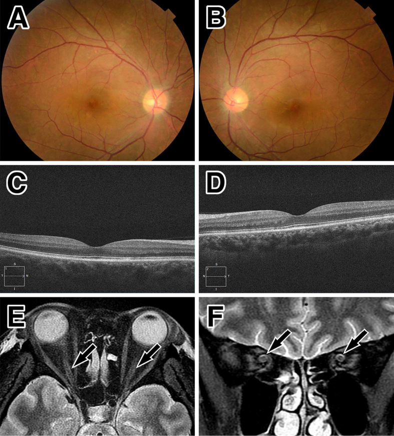
Ocular and MRI findings at the second remission. Improvement in the findings of optic disc swelling by optic fundoscopy (A: right, B: left). No subretinal fluid on optical coherence tomography at the macula (C: right, D: left), and a reduced high signal intensity (arrows) around the optic nerve on orbital MRI with short tau inversion recovery sequences in the axial (E) and coronal sections (F).