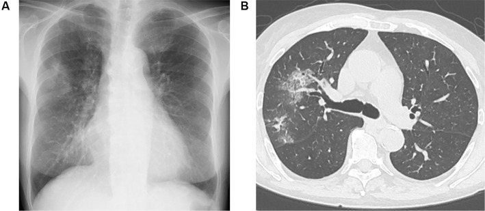
Figure 2.
(A) Chest X-ray and (B) representative axial image of the chest computed tomography before the administration of rituximab. A chest X-ray film showed the cardiothoracic ratio as 58%, and bilateral multiple patchy and ground glass opacities in the upper field of the lung. A chest computed tomographic image showed alveolar infiltrates of S2 and S3 of right lung.

