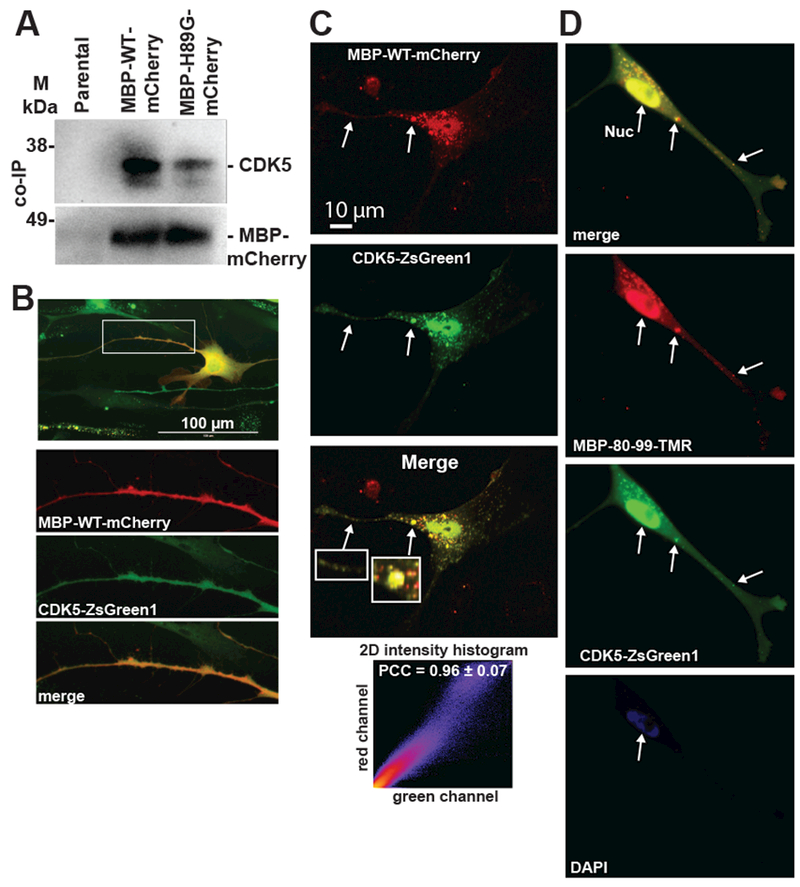
Fig. 4. MBP peptide-CDK5 Interactions.

(A) Immunoprecipitation of endogenous CDK5 in Schwann cells expressing the MBP-WT-mCherry and MBP-H89G-mCherry constructs. Proteins were co-immunoprecipitated using a mCherry antibody and analyzed by immunoblotting using anti-CDK5 and anti-mCherry antibodies. Parental Schwann cells were used as a control. M – pre-stained protein marker. (B,C) Schwann cells co-expressing the MBP-WT-mCherry (red) and CDK5-ZsGreen1 (green) constructs. (B) Enlarged images of red, green and merged channels correspond to a Schwann cell protrusion indicated by a rectangle. (C) Fluorescence intensity histogram and Pearson’s correlation coefficient (PCC) indicate the level of co-localization of the constructs in cell protrusions (5 areas). Arrows indicate the overlapping signals. (D) MBP80-99-TMR (10 μM, red) was extracellularly administered to Schwann cells expressing the CDK5-ZsGreen1 (green) construct and incubated for 1h. Overlapping signals are marked by arrows. DAPI, blue.
