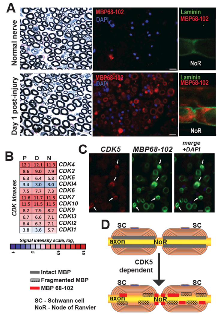
Fig. 8. Localization of CDK5 and algesic MBP peptides in PNS.

(A) Laminin (green) and MBP (MBP68-102) (red) dual-immunostaining in teased fibers, distal segment, at day 1 post-axotomy. NoR, node of Ranvier. DAPI (blue). (B) Expression of the CDK family genes in the proximal (P) and distal (D) segments of the rat sciatic nerve at day 1 post-axotomy. Heatmap represents normalized intensity values (p<0.05). Color inset shows the signal intensity log2 scale. N, intact nerve. (C) Dual-immunostaining of CDK5 (red) and MBP (MBP68-102) (green) in distal nerve sections at day 1 post-axotomy. Arrows indicate sites of co-localization. Blue, DAPI. (D) A hypothesis schematic of CDK5 dependent distribution of algesic MBP peptides in PNS. SC, Schwann cells, NoR, node of Ranvier. Intact and fragmented MBP, and MBP68-102 are indicated by gray, dotted and red rectangles, respectively.
