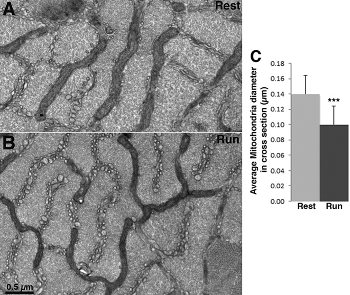
Fig. 1.
Mitochondria are smaller in fast-twitch fibers of mice at the end of a run. (A,B) Cross section of IIB/IIX fibers at the level of the I band in EDL at rest and after mice run on a treadmill to the point of exhaustion. Mitochondria are located between myofibrils in two transverse planes on either side of the Z line. Mitochondria have the same overall distribution, but they are noticeably thinner in fibers of mice after treadmill exercise (B) than in fibers of mice at rest (A). (C) Mean±s.d. mitochondrial width measured at three spots for each mitochondrion at right angle to the long axis of the organelle is 0.10±0.02 µm in fibers from exercised mice (Run) (n=251 mitochondria, 20 cells, 41 images at 26,300×, 4 mice) and 0.14±0.02 µm in fibers from resting mice (Rest) (n=190 mitochondria, 23 cells, 40 areas at 26,300×, 4 mice); Student's t-test, ***P<0.0001.

