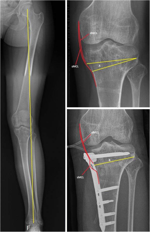
Fig. 1.

Preoperative and postoperative radiographs of the osteotomy site in relation to the medial collateral ligament. Subscript: The left figure shows a pre-operative long-leg radiographs with valgus malalignment. The right figures show a preoperative (above) and postoperative (below) radiograph of the knee. The osteotomy site (A and B) is below the insertion of the deep medial collateral ligament (dMCL). In the postoperative situation a pseudo laxity of the superficial medial collateral ligament (sMCL) is created
