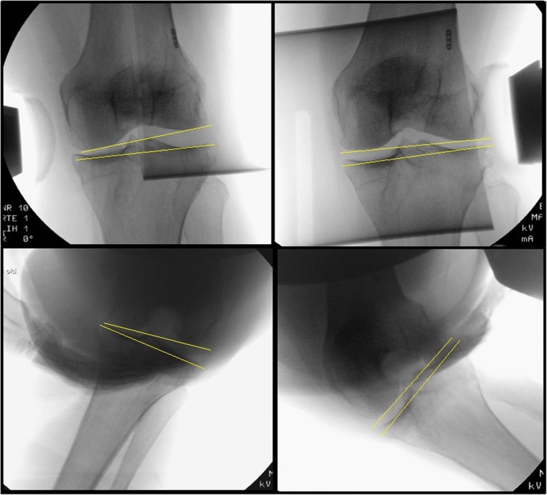
Fig. 2.

Valgus and varus laxity stress radiographs with joint space opening. Subscript: Pre-operative stress radiographs of the knee in 30° and 70° of knee flexion in an anaesthetized patient. The upper two figures show radiographs in 30° of flexion with varus (left) and valgus (right) stress. The lower two figures show radiographs in 70° of flexion in varus (left) and valgus (right) stress. The angle between a tangent line on the femur condyles and a line through the deepest tibial joint surfaces was determined and compared to the natural (unstressed) knee joint line congruence angle
