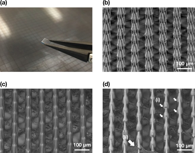
Figure 2.
(a) Photograph of a 4 × 4 mm Nanopatch held with tweezers in front of a Nanopatch wafer. (b) Representative image of secondary electron (SE) detector SEM of uncoated Nanopatch projections at a 45° tilt. (c) Representative image of backscatter electron (BSE) detector SEM of a coated Nanopatch. (d) Representative BSE detector SEM of a coated Nanopatch applied on full thickness ex vivo human skin and removed. (i) Dissolved coating representing the extent of skin penetration, and (ii) skin fragments lodged on microprojections are indicated as shown. Images shown in (a–d) are different patches but from the same wafer.

