FIGURE 4.

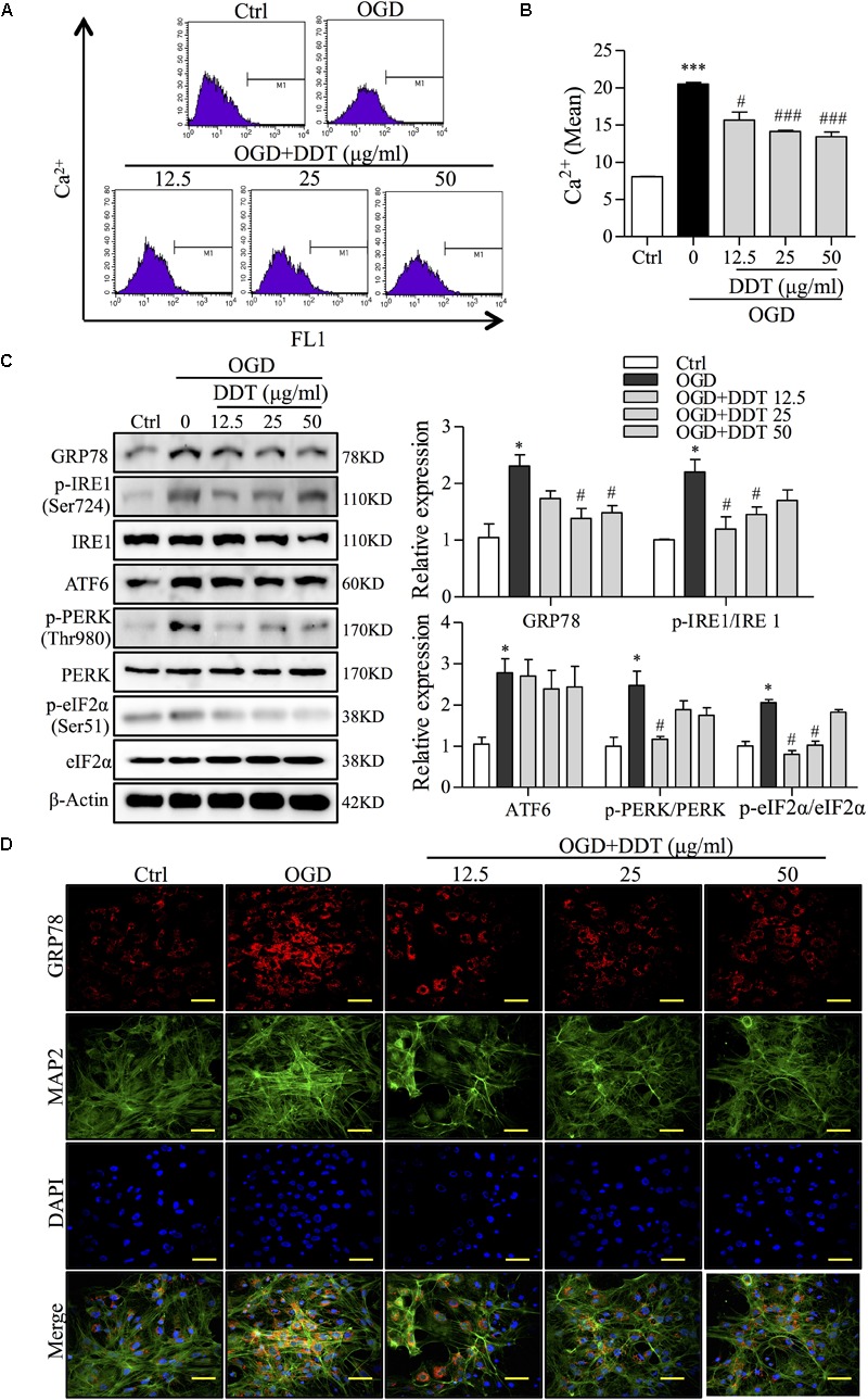
DiDang Tang inhibits Ca2+ overload and ER stress in NGF-induced PC12 cells and primary culture neurons subjected to OGD. (A) Intracellular Ca2+ levels were evaluated by FCM using the fluorescent Ca2+ indicator, Flou4-AM. (B) Bar graph represents the fluorescence intensity of Flou4-AM. (C) PC12 cells after the treatment of DDT for 48 h and an OGD incubation for 1.5 h were prepared as described in Materials and Methods and resolved by SDS–PAGE followed by Western blot using antibodies against GRP78, p-IRE1 (Ser724), IRE1, ATF6, p-PERK (Thr980), PREK, p-eIf2α (Ser51), eIf2α, and β-Actin. Relative expression was quantified with Image J and is shown on the right. (D) The expressions of GRP78 and MAP2 in primary neurons treated with DDT at different concentrations for 48 h, followed by OGD for 8 h were detected by immunofluorescence staining. Nuclei were stained with DAPI. Scale bar = 50 μm. ∗P < 0.05 and ∗∗∗P < 0.001 vs. Ctrl group; #P < 0.05 and ###P < 0.001 vs. OGD group (n = 3).
