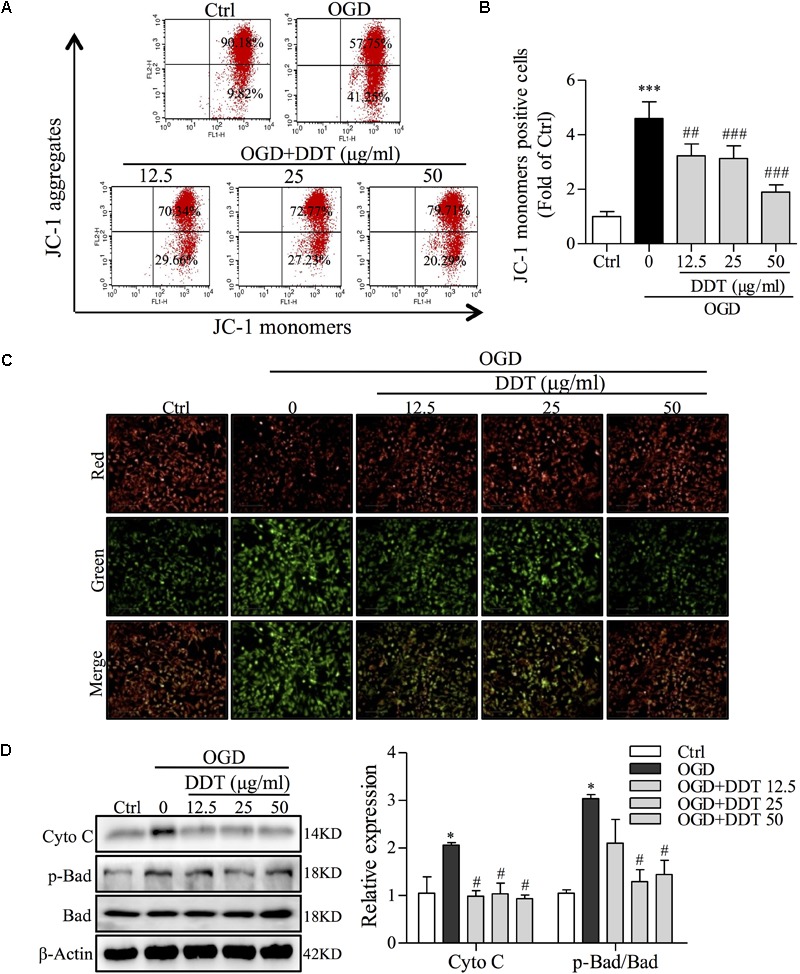
FIGURE 5.

Effects of DDT on OGD-induced intracellular mitochondrial damage in NGF-induced PC12 cells. (A) After treatment with DDT prior to OGD for 1.5 h, PC12 cells were incubated with a JC-1 probe to assess mitochondrial membrane potential (MMP) by FCM. (B) Bar graph represents the cells positive for JC-1 monomer analyzed from (A). (C) MMP of PC12 cells treated with DDT for 48 h and incubated with OGD for 1.5 h was monitored by measuring fluorescence intensity. (D) After pretreatment with DDT at different concentrations for 48 h and OGD incubation for 1.5 h, the levels of Cyto C, p-Bad and Bad in PC12 cells were detected by Western blot. ∗P < 0.05 and ∗∗∗P < 0.001 vs. Ctrl group; #P < 0.05, ##P < 0.01, and ###P < 0.001 vs. OGD group (n = 3).
