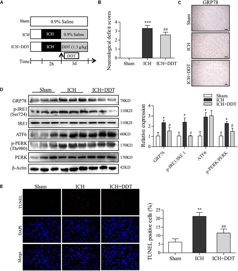
FIGURE 6.

DiDang Tang inhibits ER stress induced by ICH in a rat model. (A) Diagram of the animal study for the ICH model or the DDT treatment groups. (B) Neurological severity scores of rats in different experimental groups as described in Materials and Methods. (C) IHC staining results showed that DDT treatment inhibited the expression of GRP78 in the brain tissues around the bleeding area, compared with the ICH group. (D) The ER stress-related proteins in brain tissues from Sham, ICH, and ICH + DDT groups were analyzed by Western blot using antibodies against GRP78, p-IRE1 (Ser724), IRE1, ATF6, p-PERK (Thr980) and PREK. β-Actin was a loading control. Relative expression was quantified with Image J and is shown on the right. (E) Apoptotic cells within brain tissues were evaluated by TUNEL assays. Nuclei are counterstained with DAPI (blue). DDT treatment significantly decreased the number of apoptotic cells compared with the ICH group. Quantification of TUNEL-positive cells is shown on the right. ∗P < 0.05 and ∗∗∗P < 0.001 vs. Sham group; #P < 0.05 and ##P < 0.01 vs. ICH group (n = 5).
