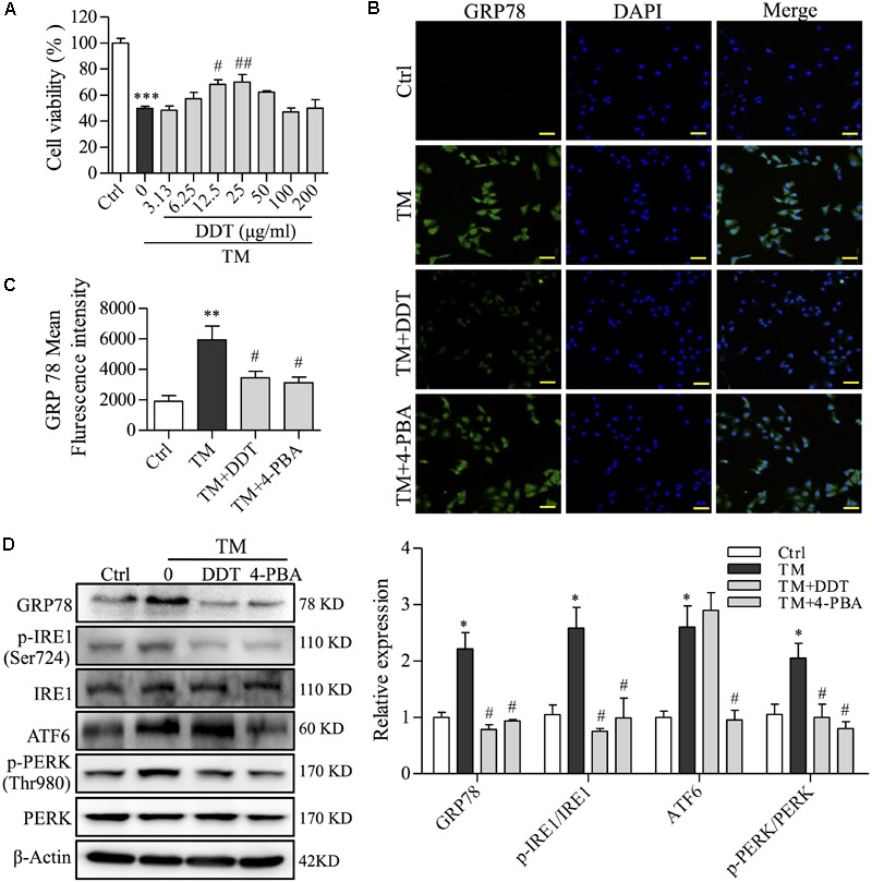
FIGURE 7.

DiDang Tang inhibits TM-induced ER stress damage in NGF-induced PC12 cells. (A) The viability of PC12 cells treated with DDT following TM incubation was determined by an MTT assay. (B) PC12 cells were treated with DDT or 4-PBA for 48 h, followed by TM incubation, and were stained with GRP78. Nuclei were stained with DAPI. Scale bar = 50 μm. (C) The bar graph represents fluorescence intensity of GRP78 expression from (B). (D) PC12 cells were treated with DDT or 4-PBA for 48 h prior to TM incubation. The levels of GRP78, p-IRE1, IRE1, ATF6, p-PERK, and PERK were examined by Western blot, and their relative expression was quantified by the means of densitometry analyses on the right. β-Actin was a loading control. An ER stress inhibitor, 4-PBA (250 μM) was used as a positive control. ∗P < 0.05, ∗∗P < 0.01, and ∗∗∗P < 0.001 vs. Ctrl group; #P < 0.05 and ##P < 0.01 vs. TM group (n = 3).
