FIGURE 8.

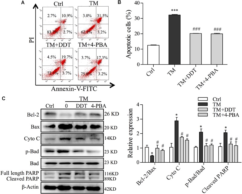
DiDang Tang inhibits ER stress-induced mitochondrial apoptosis in NGF-induced PC12 cells subjected to TM incubation. (A) After pretreatment with DDT or 4-PBA, followed by TM incubation, PC12 cells were stained with Annexin V-FITC/PI. We then detected the percentage of apoptotic cells by FCM. (B) Bar graph represents the number of apoptotic cells from (A) after the pretreatment with DDT or 4-PBA, followed by TM incubation. (C) Protein levels of Bcl-2, Bax, Cyto C, p-Bad, Bad and PARP were determined by Western blot. After the quantification by Image J, the intensity of each protein was normalized to the intensity of β-Actin and is shown on the right. ∗P < 0.05 and ∗∗∗P < 0.001 vs. Ctrl group; #P < 0.05 and ###P < 0.001 vs. TM group (n = 3).
