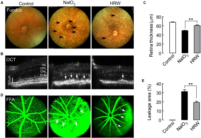
FIGURE 1.
Effects of HRW on NaIO3-induced fundus and retinal impairment in mice. After cotreatment with HRW (0.1 ml/g/day, i.g.) and NaIO3 (20 mg/kg, i.v.) for 12 days, retinas were evaluated with the fundus photography, OCT examination, and FFA examination. (A) Representative images of fundus photography. The black arrow represents yellow-white, drusen-like structures. (B) Representative images of OCT in the mouse retina and (C) quantification of retinal thickness. White arrows indicate areas of hyperreflexia in the RPE area. (D) Representative images of FFA and (E) quantification of the leakage area. White arrowheads represent the leaking area. RPE, retinal pigment epithelium; IS/OS, inner segment/outer segment; ONL, outer nuclear layer; OPL, outer plexiform layer; INL, inner nuclear layer. Values are presented as the mean ± SEM, n = 5, ∗∗P < 0. 01 vs. the NaIO3 group.

