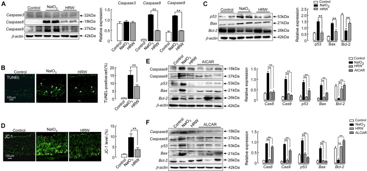
FIGURE 6.
HRW reduces apoptosis in the retina by upregulating Sirt1. (A) Representative blot and quantitative analysis of caspase 3, caspase 8, and caspase 9 protein expression levels at 5 days after NaIO3 injection with or without HRW administration. (B) Representative images of TUNEL-stained retinal sections from control and NaIO3 mice with or without HRW administration. TUNEL-positive cells are shown by asterisks. Data represent the mean ± SEM of three independent experiments. n = 10. (C) Representative blot and the quantitative analysis of protein expression for p53, Bax, and Bcl-2 expression in the retina. n = 4. (D) Representative images of Jc-1-stained retinal sections and the quantification of green fluorescence intensity in retinal sections. n = 5. Representative Western blot and the quantitative analysis of caspase 8, caspase 9, p53, Bax, and Bcl-2 protein expression after ALCAR (E) or AICAR (F) injection in retinas from NaIO3 mice. Values were presented as the mean ± SEM. n = 3–6, ∗∗P < 0.01 vs. the NaIO3 group.

