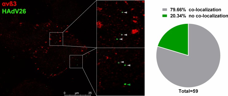
FIG 17.
Colocalization of Alexa Fluor 488-labeled HAdV26 with αvβ3 integrin in A549-B6 cells. Cells were incubated with Alexa Fluor 488-labeled HAdV26 (50,000 vp/cell) for 1 min at 37°C, fixed with 2% PFA, and subsequently stained for αvβ3 integrin expression (LM609). A representative confocal image of HAdV26 colocalized with αvβ3 integrin is shown. The gray arrowheads indicate colocalization; the green arrowhead indicates absence of colocalization. The pie chart represents quantification of the percentage of colocalized HAdV26 with αvβ3 integrin. The data were collected from 9 cells and 59 viruses that infected the cells.

