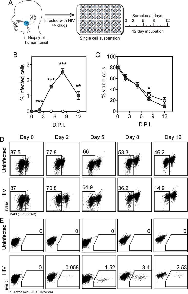
FIG 2.

Human lymphoid aggregate culture (HLAC) of tonsil explant model supports HIV-1 infection. (A) Human tonsil explants were collected, dissected, homogenized, and passed through a cell strainer. Cells were subjected to Ficoll fractionation, and human lymphoid aggregate cells (HLACs) were plated and then infected with HIV-1 NL-CI. Cells were collected at 0, 2, 5, 8, and 12 dpi. (Printed with permission from Mount Sinai Health System.) (B) HLACs were collected on the indicated days and analyzed by flow cytometry for productive infection by NL-CI mCherry fluorescence. Infected cells were quantified by the percentage of positive PE-Texas Red events. (C) HLACs were analyzed by flow cytometry to quantify viable cells. Viable cells were quantified by the percentage of negative DAPI events. (D) Representative flow cytometry plots of uninfected and infected cells are shown for Live/Dead Fixable Dead Cell staining (Thermo Fisher) by flow cytometry, indicating viability of the 12-day infection time course. (D) Representative flow cytometry plots of uninfected and infected cells are shown for mCherry signal as indicative of HIV-1 productive infection. Mean values ± standard errors of the means are presented from three donors. *, P ≤ 0.05; **, P ≤ 0.01; ***, P ≤ 0.001.
