FIGURE 4.

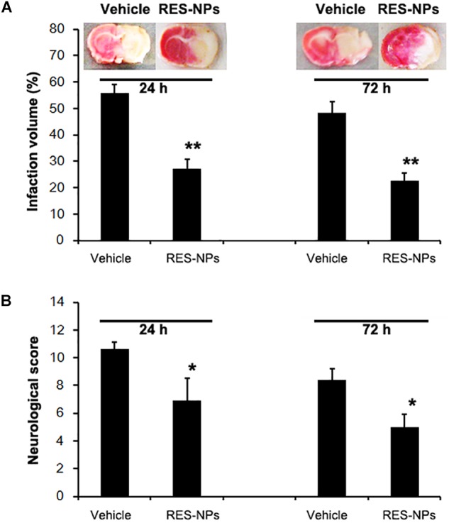
Reduction of neurological score and infarct volume by RES-HSA-NPs (RES-NPs) in rats suffering ischemia-reperfusion. At the onset of reperfusion following 2 h of ischemia, rats received via tail vein either vehicle (control) or a suspension of 20 mg/kg of RES-HSA-NPs. Animals were euthanized 24 and 72 h following reperfusion, respectively. (A) Representative coronal brain sections stained with TTC solution from vehicle or RES-NPs treated rats. Red colored regions in the TTC-stained sections indicate non-ischemic areas; pale-colored regions indicate ischemic portions. Bar graph showing quantification of infarction volume from the vehicle and RES-NPs treated rats. (B) Bar graph showing quantification of infarction volume from the vehicle and RES-NPs treated rats. Data are means ± SD. ∗P < 0.05 vs. PBS control, ∗∗P < 0.01 vs. vehicle (n = 5 per group).
