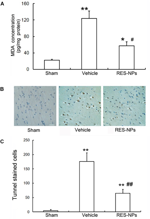
FIGURE 5.

Inhibition of oxidative stress and apoptosis by RES-HSA-NPs (RES-NPs) in rats following ischemia-reperfusion. (A) RES-NPs attenuated oxidative stress in brain after I/R. Quantified (n = 4) MDA levels of the cerebral cortex from I/R side after 24 h reperfusion. The RES-NPs (20 mg/kg) treatment had significantly decreased cortex MDA levels compared with the vechicle (∗∗P < 0.01 vs. sham, ##P < 0.01 vs. vehicle, ANOVA). (B) Representative photomicrographs showing yellow-brown TUNEL staining cells (pointed by white arrows) in penumbral region of brain sections from Sham, vehicle and RES-NPs treated rats after 72 h reperfusion (n = 4). Scale bars = 100 μm. (C) Quantification of the TUNEL-staining cells. ∗P < 0.05 vs. sham, #P < 0.05 vs. vehicle.
