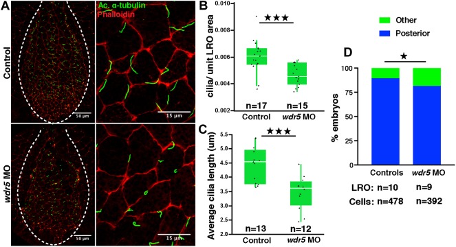
Fig. 2.
Wdr5 depletion affects cilia morphology in the LRO of X.tropicalis. (A) Cilia in control and wdr5 morphant Xenopus LROs marked by anti-acetylated (Ac.) α-tubulin (green) antibody and F-actin marked by phalloidin (red). (B,C) Number of cilia normalized to the LRO area (B) and length of cilia (C) in uninjected controls and wdr5 morphants. Data are presented as box plot with 95% confidence interval. ‘n’=number of embryos. (D) Number of cilia posteriorly localized in the LRO cells in uninjected controls and wdr5 morphants. Experiments were repeated two times. ★P<0.05 and ★★★P<0.0005.

